


AI in medical imaging: Transforming medical image analysis


May 6, 2026 10 min read 84 views
One of the biggest areas of innovation in healthcare is currently artificial intelligence, and one of the leading areas of that movement is medical imaging. Hospitals, researchers, and medtech organizations are leveraging deep learning algorithms trained on large image datasets to support image classification, triage of urgent scans, identification of subtle abnormalities, and reduction of repetitive tasks associated with image reading during normal daily patient care. Thus, the development of AI for imaging is no longer a niche effort; instead, it is becoming an integral part of the evolution process of contemporary diagnostic workflows.
A well-established example is Mayo Clinic. They use artificial intelligence in radiology to help locate tumors and structures, assess body composition on CT imaging, and support the detection of findings such as intracranial aneurysms, strokes, or pulmonary embolisms. This example gives you a good idea of which direction the market will take, so the following sections will provide more specifics as to how this will work.
Medical imaging technology key takeaways
- Advanced AI has transitioned from experimentation to practical use across multiple types of medical imaging, supporting the detection, triage, segmentation, and workflow prioritization of images.
- The most useful applications of AI in medical imaging are as assistive technologies for clinicians—intended to help them work more quickly and consistently—rather than replacing their clinical judgment.
- Current tools have the greatest impact on high-volume, pattern-based tasks such as screening, early detection, and urgent-case triage.
- Ongoing AI research in healthcare continues to explore the clinical value of AI; it will rely on the model’s accuracy but also on other aspects, including generalizability, control of bias, suitability for the clinical workflow, and real-world adoption.
The current state of AI in medical imaging and radiology
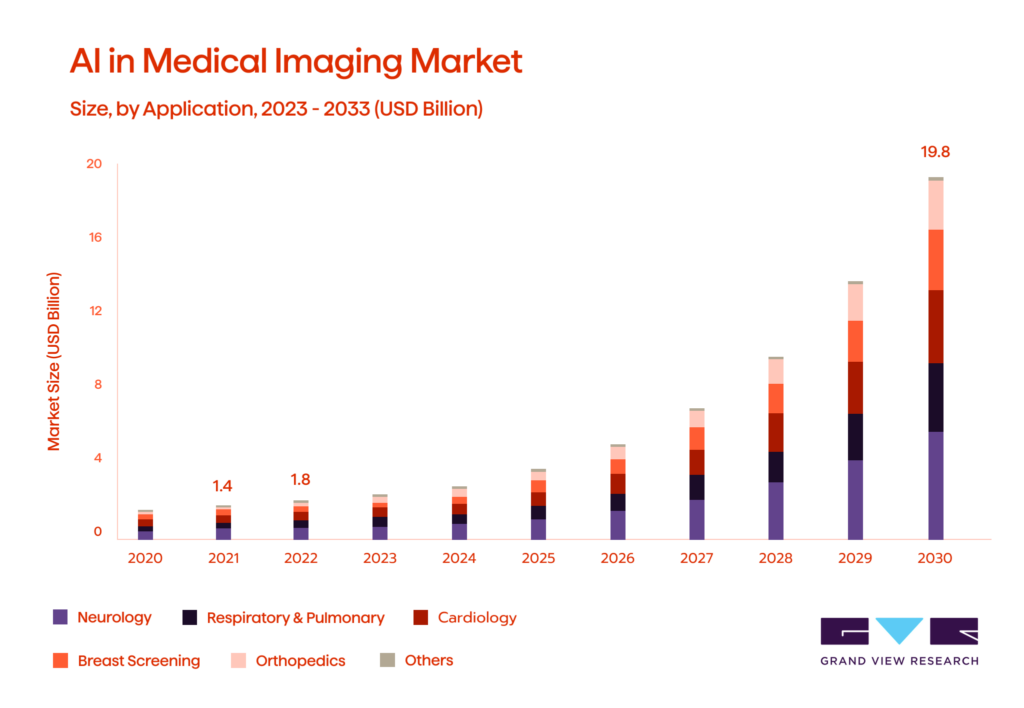
The data clearly indicates the direction the market is headed. According to estimates, the global AI in medical imaging market was $1.36 billion in 2024 and is expected to reach $19.78 billion by 2033, representing a CAGR of 34.67% from 2025 to 2033. This demonstrates how rapidly AI technologies are being adopted into everyday radiology practice, having been heavily developed/explored through research. In fact, hospitals and vendors have moved beyond feasibility studies. They are now developing products for triage, lesion detection, segmentation, quantification, report support, and workflow prioritization of the increasing volume of imaging data.
Radiology is leading the shift towards clinical AI tools. According to a 2025 policy review, over 75% of FDA-approved clinical AI tools were used for Radiology. The FDA-listed AI-enabled devices also show that Radiology remains the most prominent specialty. AI adoption is also increasing on the clinical side, as evidenced by a 2024 survey of Radiologists in Europe, where 48% reported already using AI tools, while 25% are (or were) planning to implement them. This is a more accurate perspective on AI development in Imaging: the novelty stage is complete; however, we are still in a transition phase from pilot implementation to full operational maturity.
AI performs best when performing certain tasks repeatedly for many patients. A multicenter example is that a radiologist who detected cancers with an AI-CAD program (computer-aided detection) did so more often than radiologists reading mammograms without AI-CAD. This shows the impact of machine learning on the early detection of breast cancer by augmenting radiologists rather than acting independently in the diagnostic process.
The cautious nature of the AI industry stems from current experience with broad-based multi-model applications that struggle to interpret X-rays. As a result of this new technology, with the potential for efficiency, consistency, and partial assistance in treatment planning, the degree to which clinicians trust newly developed AI applications will not only be influenced by the AI model’s performance, but also by the governing bodies responsible for approving these applications for clinical use and how the newly developed application fits into the existing clinical workflow.
Enhance research capabilities and optimize Ru0026amp;D strategies with tailored digital technology.
Deep learning in radiology: from detection support to diagnostic decision-making
AI has become increasingly integrated into healthcare today, and “deep learning” is a big part of that. Rather than being guided by explicit rules (hand-crafted rules) like prior artificial intelligence systems, deep learning models use imaging data as input to learn patterns directly out of an image file. This is particularly beneficial for radiology and the analysis of medical images, as the visual criteria that may differentiate one diagnosis from another can be subtle yet have a significant impact on how the diagnosis is treated.
Deep learning is currently utilized for diagnostic purposes. For example, deep learning will provide a second set of eyes to help detect potential lung nodules on CT scans, identify possible fractures on X-ray images, segment tumors on MRIs, and rank high-priority results in a triage list. Deep learning reduces the risk of human error, allows the radiologist to review cases more efficiently, and directs their attention to the area of greatest need.
Moving forward, AI will provide additional support in the decision-making process beyond just identifying abnormalities on imaging tests. For example, AI can estimate the probability of malignancy for a particular abnormality, compare imaging findings with previously identified similar abnormalities, calculate the total number of abnormalities in a given patient, and suggest the most likely alternative causes for the abnormalities identified on the imaging test. While full automation of these tasks is unlikely in most cases, radiologists may rely on AI to aid their decision-making.
How AI-powered image analysis works across imaging modalities
Once deep learning has been integrated into diagnostic decision-making, determining how it will actually work depends heavily upon the type of scan. The type of scan determines how a signal is produced, the type of structure it contains, and, lastly, the clinical pattern it reveals.
AI applications in CT and MRI include image segmentation, lesion detection, volumetric measurement, tumor volume calculation, and tracking disease progression, using a range of models. They can delineate tumors, organs, hemorrhages, or vascular structures much faster than human reviewers can, particularly beneficial in oncology, neurology, and emergency medicine.
X-ray analysis typically has two main focus areas: classification and triage. The primary objective of AI tools is to accurately detect urgent transient pathologies, such as fractures, pneumothorax, pneumonia, and other urgent abnormalities, in high-throughput environments where speed is critical. Ultrasound is very dynamic, so AI typically assists with selecting the correct frame, labeling the anatomy, and providing measurement assistance during live exams.
AI is used in digital pathology and many other forms of biomedical imaging systems. AI applications identify patterns in tissue, cell shapes, and tumor characteristics from whole slide images. The speed at which AI can process this information is a key factor in advancing AI technologies beyond radiology into other areas of medicine. The same basic idea holds across the various modalities in which AI operates: converting an unstructured image into a structured clinical signal. However, each modality has a different workflow and a different level of decision-support requirement.
Early detection, triage, and screening use cases in AI in medical imaging
The medical field has significant potential for AI implementation at the point of patient care and diagnosis. In addition, early screening/triage/detection identifies many patients with a pattern in their data. Therefore, this makes them a very applicable solution for using deep learning-based models in this area. Although it is not intended to replace physician judgment, AI solutions generally help identify urgent cases earlier, reduce the workload to interpret results, and standardize how screening processes are performed.
The most significant advancements in early detection are in cancer screening. AI is currently used in mammography to help determine which exams are likely normal, mark those that appear suspicious, and assist radiologists in detecting cancer while reducing workload. Similar developments are underway in ultrasound screening, as studies have demonstrated improved accuracy in AI-assisted liver imaging workflows while reducing radiologists’ workload. Due to this experience, current AI system development supporting the screening process focuses as much on the workflow’s impact as on the individual model’s accuracy.
Triage is another use case for AI in radiology. This time, however, the unique use case of AI is not to generate a final report first. Rather, it examines the workload list, assigns priorities based on time, and puts findings for which time is of the essence first. This is particularly important in brain imaging for a stroke or intracranial hemorrhage and chest CTs looking for pulmonary embolisms, where a few minutes could change the overall outcome of a patient. There are FDA-cleared tools already available for these types of radiology triaging scenarios.
| Use case | Typical modality | What AI helps with | Clinical value |
| Early detection | Mammography, ultrasound | Finds subtle suspicious patterns | Earlier intervention |
| Triage | CT, especially brain and chest imaging | Prioritizes urgent studies | Faster review of critical cases |
| Screening | Breast, liver, lung imaging | Separates likely normal from higher-risk exams | Lower workload, more scalable medical practice |
How artificial intelligence in medical imaging can improve patient outcomes
The clearest value of AI in imaging is not replacing clinical staff, but speeding up the care process by reducing missed findings and improving consistency in provider decision-making. In practice, through the adoption of AI, radiologists can identify and act on abnormal scans more quickly than before; identify and reliably detect small abnormalities more consistently than before; and perform their job duties at a higher volume than before. This is important because delayed diagnostics have a ripple effect on patient triage, specialist consultation, and treatment timing.
AI-assisted reading in breast imaging has improved breast cancer detection, as demonstrated in numerous prospective studies. In acute stroke and, less commonly, for hemorrhage or other emergency care settings, AI triage tools expedite the delivery of critical imaging studies to operators, which can be critical when treatment windows are narrow.
In outcome terms, the use of AI can support:
- earlier detection of clinically important findings
- faster escalation of time-sensitive cases
- more consistent interpretation across readers
- better measurement of disease burden on diagnostic imaging
- lower workload for repetitive, high-volume tasks
AI models do not add value in isolation; they enhance clinical decision-making. In that way, medical imaging applications are most valuable when they improve timeliness, consistency, and diagnostic confidence in patient care. Therefore, AI has the opportunity to make technical performance translate into better patient care.
FAQ
Embracing medical AI systems with Avenga
The way AI will support clinicians in medical imaging is trending toward a more collaborative model, enabling them to analyze images more rapidly, prioritize findings more straightforwardly, and interpret them consistently. The ultimate advantage of AI in medical imaging is not simply the automation of the imaging process, but also its ability to help radiologists and care teams make sound clinical decisions under pressure.
Want to learn more about AI and medical imaging? Contact Avenga, your trusted AI imaging partner.


Your business results matter
Achieve them with minimized risk through our bespoke innovation capabilities. Fill in the form below.